Abstract
Case Report
Hepatic adenomatosis: A clinically challenging rare liver disease
Sunitha Ramachandra*, Lakshmi Rao and Masoud Al Kindi
Published: 11 July, 2018 | Volume 2 - Issue 1 | Pages: 005-008
43-year-old lady presented with incidentally discovered liver lesions while she was being managed for her complaints of menorrhagia. CT and MRI showed hepatomegaly with multiple lesions in both lobes of the liver with vascular element in the background of diffuse fatty infiltration. Patient underwent laparoscopic core biopsy. Histopathology showed extensive steatosis, intracytoplasmic giant mitochondria and absence of portal tracts, features highly suggestive of hepatic adenomatosis. IHC staining showed membranous and cytoplasmic positivity in hepatocytes for B-catenin consistent with multiple hepatic adenomatosis. Hepatic adenomatosis is a new clinical entity in the hepatological practice characterized by the presence of 10 or more nodules in the liver known for its major complication of bleeding. Hepatic adenomatosis is managed by regular imaging and resection of large (> 5cm) superficial and painful adenomas along with liver function tests and tumor markers to rule out malignant transformation. However, the potential cure being the liver transplantation.
Read Full Article HTML DOI: 10.29328/journal.acgh.1001006 Cite this Article Read Full Article PDF
References
- Barthelmes L, Tait I. Liver cell adenoma and liver cell adenomatosis. HPB. 2005; 7:186-196. Ref.: https://tinyurl.com/yboxbcdc
- Donato M, Jahromi AH, Andrade A, Kim R, Chaudhery SI, et al. Hepatic Adenomatosis: A rare but important liver disease with severe clinical implications. Int Surg. 2015; 100: 903-907. Ref.: https://tinyurl.com/ybhb6xbr
- Geller SA, Petrovic LD. Tumour and tumour-like conditions. In: Biopsy Interpretation of the Liver. 2nd edition. Philadelphia: Wolters Kluwer/Lippincott Williams & Wilkins. 2009; 318.
- Chiche L, Dao T, Salamé E, Galais MP, Bouvard N, et al. Liver Adenomatosis: Reappraisal, Diagnosis, and Surgical Management: Eight New Cases and Review of the Literature. Ann Surg. 2000; 231: 74-81. Ref.: https://tinyurl.com/ybgbxrc6
Figures:

Figure 1
Figure 2
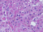
Figure 3
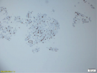
Figure 4
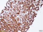
Figure 5
Similar Articles
-
A rare cause of obstructive jaundice - case reportPriya Mohan*,Sumathi Bavanandam,Sunil Kumar KS. A rare cause of obstructive jaundice - case report . . 2017 doi: 10.29328/journal.hcg.1001001; 1: 001-003
-
Anemia due to a rare anomaly - Case ReportS Nanthakumar*,Sumathi Bavanandam,Nirmala Dheivamani,Natarajan B, Krishna Mohan. Anemia due to a rare anomaly - Case Report . . 2017 doi: 10.29328/journal.hcg.1001002; 1: 004-006
-
The influence of Infliximab on the development of Experimental PancreatitisLychkova AE*,Golubev Yu Yu, Puzikov AM. The influence of Infliximab on the development of Experimental Pancreatitis . . 2017 doi: 10.29328/journal.hcg.1001003; 1: 007-011
-
Role of Accessory Right Inferior Hepatic Veins in evaluation of Liver TransplantationAwais Ahmed,Abdu Hafeez-Baig*, Mirza Akmal Sharif,Umair Ahmed,Raj Gururajan. Role of Accessory Right Inferior Hepatic Veins in evaluation of Liver Transplantation . . 2017 doi: 10.29328/journal.acgh.1001004; 1: 012-016
-
Analysis of Pyogenic Liver AbscessesEva Barreiro Alonso**. Analysis of Pyogenic Liver Abscesses . . 2018 doi: 10.29328/journal.acgh.1001005; 2: 001-004
-
Hepatic adenomatosis: A clinically challenging rare liver diseaseSunitha Ramachandra*,Lakshmi Rao, Masoud Al Kindi . Hepatic adenomatosis: A clinically challenging rare liver disease. . 2018 doi: 10.29328/journal.acgh.1001006; 2: 005-008
-
An uncommon cause of isolated ascites: Pseudomyxoma peritoneiLouly Hady*,I Nassar, K Znati,N Kabbaj. An uncommon cause of isolated ascites: Pseudomyxoma peritonei . . 2019 doi: 10.29328/journal.acgh.1001007; 3: 001-005
-
Transcatheter Arterial Embolization for the treatment of upper gastrointestinal bleedingMohammed Habib*,Majed Alshounat. Transcatheter Arterial Embolization for the treatment of upper gastrointestinal bleeding . . 2019 doi: 10.29328/journal.acgh.1001008; 3: 006-011
-
Endoscopic treatment of pancreatic diseases via Duodenal Minor Papilla: 135 cases treated by Sphincterotomy, Endoscopic Pancreatic Duct Balloon Dilation (EPDBD), and Pancreatic Stenting (EPS)Tadao Tsuji*, G Sun,A Sugiyama, Y Amano,S Mano,T Shinobi,H Tanaka,M Kubochi,K Ohishi,Y Moriya,M Ono,T Masuda, H Shinozaki,H Kaneda,H Katsura,T Mizutani, K Miura,M Katoh, K Yamafuji, K Takeshima,N Okamoto,Y Hoshino,N Tsurumi,S Hisada,J Won,T Kogiso,K Yatsuji,M Iimura, T Kakimoto,S Nyuhzuki. Endoscopic treatment of pancreatic diseases via Duodenal Minor Papilla: 135 cases treated by Sphincterotomy, Endoscopic Pancreatic Duct Balloon Dilation (EPDBD), and Pancreatic Stenting (EPS) . . 2019 doi: 10.29328/journal.acgh.1001009; 3: 012-019
-
Addition of Simvastatin to Carvedilol and Endoscopic Variceal Ligation improves rebleeding and survival in patients with Child-Pugh A and B class but not in Child Pugh C classSanjeev Kumar Jha*,Kuldeep Saharawat,Ravi Keshari,,Praveen Jha,Shubham Purkayastha, Ravish Ranjan . Addition of Simvastatin to Carvedilol and Endoscopic Variceal Ligation improves rebleeding and survival in patients with Child-Pugh A and B class but not in Child Pugh C class . . 2019 doi: 10.29328/journal.acgh.1001010; 3: 020-026
Recently Viewed
-
Cystoid Macular Oedema Secondary to Bimatoprost in a Patient with Primary Open Angle GlaucomaKonstantinos Kyratzoglou*,Katie Morton. Cystoid Macular Oedema Secondary to Bimatoprost in a Patient with Primary Open Angle Glaucoma. Int J Clin Exp Ophthalmol. 2025: doi: 10.29328/journal.ijceo.1001059; 9: 001-003
-
Navigating Neurodegenerative Disorders: A Comprehensive Review of Current and Emerging Therapies for Neurodegenerative DisordersShashikant Kharat*, Sanjana Mali*, Gayatri Korade, Rakhi Gaykar. Navigating Neurodegenerative Disorders: A Comprehensive Review of Current and Emerging Therapies for Neurodegenerative Disorders. J Neurosci Neurol Disord. 2024: doi: 10.29328/journal.jnnd.1001095; 8: 033-046
-
Obesity in Patients with Chronic Obstructive Pulmonary Disease as a Separate Clinical PhenotypeDaria A Prokonich*, Tatiana V Saprina, Ekaterina B Bukreeva. Obesity in Patients with Chronic Obstructive Pulmonary Disease as a Separate Clinical Phenotype. J Pulmonol Respir Res. 2024: doi: 10.29328/journal.jprr.1001060; 8: 053-055
-
Current Practices for Severe Alpha-1 Antitrypsin Deficiency Associated COPD and EmphysemaMJ Nicholson*, M Seigo. Current Practices for Severe Alpha-1 Antitrypsin Deficiency Associated COPD and Emphysema. J Pulmonol Respir Res. 2024: doi: 10.29328/journal.jprr.1001058; 8: 044-047
-
Clinical and Histopathological Mismatch: A Case Report of Acral FibromyxomaMonica Mishra*,Kailas Mulsange,Gunvanti Rathod,Deepthi Konda. Clinical and Histopathological Mismatch: A Case Report of Acral Fibromyxoma. Arch Pathol Clin Res. 2025: doi: 10.29328/journal.apcr.1001045; 9: 005-007
Most Viewed
-
Evaluation of Biostimulants Based on Recovered Protein Hydrolysates from Animal By-products as Plant Growth EnhancersH Pérez-Aguilar*, M Lacruz-Asaro, F Arán-Ais. Evaluation of Biostimulants Based on Recovered Protein Hydrolysates from Animal By-products as Plant Growth Enhancers. J Plant Sci Phytopathol. 2023 doi: 10.29328/journal.jpsp.1001104; 7: 042-047
-
Sinonasal Myxoma Extending into the Orbit in a 4-Year Old: A Case PresentationJulian A Purrinos*, Ramzi Younis. Sinonasal Myxoma Extending into the Orbit in a 4-Year Old: A Case Presentation. Arch Case Rep. 2024 doi: 10.29328/journal.acr.1001099; 8: 075-077
-
Feasibility study of magnetic sensing for detecting single-neuron action potentialsDenis Tonini,Kai Wu,Renata Saha,Jian-Ping Wang*. Feasibility study of magnetic sensing for detecting single-neuron action potentials. Ann Biomed Sci Eng. 2022 doi: 10.29328/journal.abse.1001018; 6: 019-029
-
Pediatric Dysgerminoma: Unveiling a Rare Ovarian TumorFaten Limaiem*, Khalil Saffar, Ahmed Halouani. Pediatric Dysgerminoma: Unveiling a Rare Ovarian Tumor. Arch Case Rep. 2024 doi: 10.29328/journal.acr.1001087; 8: 010-013
-
Physical activity can change the physiological and psychological circumstances during COVID-19 pandemic: A narrative reviewKhashayar Maroufi*. Physical activity can change the physiological and psychological circumstances during COVID-19 pandemic: A narrative review. J Sports Med Ther. 2021 doi: 10.29328/journal.jsmt.1001051; 6: 001-007

HSPI: We're glad you're here. Please click "create a new Query" if you are a new visitor to our website and need further information from us.
If you are already a member of our network and need to keep track of any developments regarding a question you have already submitted, click "take me to my Query."


