Figure 4
Pitfalls in the hemostatic management of a liver transplantation
Raveh Yehuda* and Nicolau-Raducu Ramona
Published: 13 April, 2022 | Volume 6 - Issue 1 | Pages: 001-005
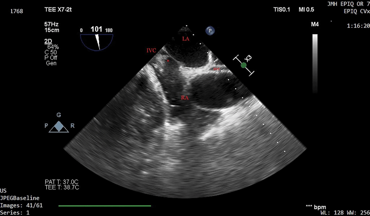
Figure 4:
Two-dimensional transesophageal echocardiographic bicaval view during a severe hypotensive episode, 10 minutes prior to reperfusion. A large thrombus is seen at the confluence of the inferior vena cava and the right atrium. Heparin 2000 units and vasopressors were administered and hemodynamic stability was restored. The clot dispersed over 10 minutes. LA, left atrium; RA, right atrium, IVC, inferior vena cava; SVC, superior vena cava; * intracardiac clot.
Read Full Article HTML DOI: 10.29328/journal.acgh.1001032 Cite this Article Read Full Article PDF
More Images
Similar Articles
-
A rare cause of obstructive jaundice - case reportPriya Mohan*,Sumathi Bavanandam,Sunil Kumar KS. A rare cause of obstructive jaundice - case report . . 2017 doi: 10.29328/journal.hcg.1001001; 1: 001-003
-
Anemia due to a rare anomaly - Case ReportS Nanthakumar*,Sumathi Bavanandam,Nirmala Dheivamani,Natarajan B, Krishna Mohan. Anemia due to a rare anomaly - Case Report . . 2017 doi: 10.29328/journal.hcg.1001002; 1: 004-006
-
The influence of Infliximab on the development of Experimental PancreatitisLychkova AE*,Golubev Yu Yu, Puzikov AM. The influence of Infliximab on the development of Experimental Pancreatitis . . 2017 doi: 10.29328/journal.hcg.1001003; 1: 007-011
-
Role of Accessory Right Inferior Hepatic Veins in evaluation of Liver TransplantationAwais Ahmed,Abdu Hafeez-Baig*, Mirza Akmal Sharif,Umair Ahmed,Raj Gururajan. Role of Accessory Right Inferior Hepatic Veins in evaluation of Liver Transplantation . . 2017 doi: 10.29328/journal.acgh.1001004; 1: 012-016
-
Analysis of Pyogenic Liver AbscessesEva Barreiro Alonso**. Analysis of Pyogenic Liver Abscesses . . 2018 doi: 10.29328/journal.acgh.1001005; 2: 001-004
-
Hepatic adenomatosis: A clinically challenging rare liver diseaseSunitha Ramachandra*,Lakshmi Rao, Masoud Al Kindi . Hepatic adenomatosis: A clinically challenging rare liver disease. . 2018 doi: 10.29328/journal.acgh.1001006; 2: 005-008
-
An uncommon cause of isolated ascites: Pseudomyxoma peritoneiLouly Hady*,I Nassar, K Znati,N Kabbaj. An uncommon cause of isolated ascites: Pseudomyxoma peritonei . . 2019 doi: 10.29328/journal.acgh.1001007; 3: 001-005
-
Transcatheter Arterial Embolization for the treatment of upper gastrointestinal bleedingMohammed Habib*,Majed Alshounat. Transcatheter Arterial Embolization for the treatment of upper gastrointestinal bleeding . . 2019 doi: 10.29328/journal.acgh.1001008; 3: 006-011
-
Endoscopic treatment of pancreatic diseases via Duodenal Minor Papilla: 135 cases treated by Sphincterotomy, Endoscopic Pancreatic Duct Balloon Dilation (EPDBD), and Pancreatic Stenting (EPS)Tadao Tsuji*, G Sun,A Sugiyama, Y Amano,S Mano,T Shinobi,H Tanaka,M Kubochi,K Ohishi,Y Moriya,M Ono,T Masuda, H Shinozaki,H Kaneda,H Katsura,T Mizutani, K Miura,M Katoh, K Yamafuji, K Takeshima,N Okamoto,Y Hoshino,N Tsurumi,S Hisada,J Won,T Kogiso,K Yatsuji,M Iimura, T Kakimoto,S Nyuhzuki. Endoscopic treatment of pancreatic diseases via Duodenal Minor Papilla: 135 cases treated by Sphincterotomy, Endoscopic Pancreatic Duct Balloon Dilation (EPDBD), and Pancreatic Stenting (EPS) . . 2019 doi: 10.29328/journal.acgh.1001009; 3: 012-019
-
Addition of Simvastatin to Carvedilol and Endoscopic Variceal Ligation improves rebleeding and survival in patients with Child-Pugh A and B class but not in Child Pugh C classSanjeev Kumar Jha*,Kuldeep Saharawat,Ravi Keshari,,Praveen Jha,Shubham Purkayastha, Ravish Ranjan . Addition of Simvastatin to Carvedilol and Endoscopic Variceal Ligation improves rebleeding and survival in patients with Child-Pugh A and B class but not in Child Pugh C class . . 2019 doi: 10.29328/journal.acgh.1001010; 3: 020-026
Recently Viewed
-
A Gateway to Metal Resistance: Bacterial Response to Heavy Metal Toxicity in the Biological EnvironmentLoai Aljerf*,Nuha AlMasri. A Gateway to Metal Resistance: Bacterial Response to Heavy Metal Toxicity in the Biological Environment. Ann Adv Chem. 2018: doi: 10.29328/journal.aac.1001012; 2: 032-044
-
Obesity in Patients with Chronic Obstructive Pulmonary Disease as a Separate Clinical PhenotypeDaria A Prokonich*, Tatiana V Saprina, Ekaterina B Bukreeva. Obesity in Patients with Chronic Obstructive Pulmonary Disease as a Separate Clinical Phenotype. J Pulmonol Respir Res. 2024: doi: 10.29328/journal.jprr.1001060; 8: 053-055
-
Current Practices for Severe Alpha-1 Antitrypsin Deficiency Associated COPD and EmphysemaMJ Nicholson*, M Seigo. Current Practices for Severe Alpha-1 Antitrypsin Deficiency Associated COPD and Emphysema. J Pulmonol Respir Res. 2024: doi: 10.29328/journal.jprr.1001058; 8: 044-047
-
Navigating Neurodegenerative Disorders: A Comprehensive Review of Current and Emerging Therapies for Neurodegenerative DisordersShashikant Kharat*, Sanjana Mali*, Gayatri Korade, Rakhi Gaykar. Navigating Neurodegenerative Disorders: A Comprehensive Review of Current and Emerging Therapies for Neurodegenerative Disorders. J Neurosci Neurol Disord. 2024: doi: 10.29328/journal.jnnd.1001095; 8: 033-046
-
Metastatic Brain Melanoma: A Rare Case with Review of LiteratureNeha Singh,Gaurav Raj,Akshay Kumar,Deepak Kumar Singh,Shivansh Dixit,Kaustubh Gupta*. Metastatic Brain Melanoma: A Rare Case with Review of Literature. J Radiol Oncol. 2025: doi: 10.29328/journal.jro.1001080; 9: 050-053
Most Viewed
-
Evaluation of Biostimulants Based on Recovered Protein Hydrolysates from Animal By-products as Plant Growth EnhancersH Pérez-Aguilar*, M Lacruz-Asaro, F Arán-Ais. Evaluation of Biostimulants Based on Recovered Protein Hydrolysates from Animal By-products as Plant Growth Enhancers. J Plant Sci Phytopathol. 2023 doi: 10.29328/journal.jpsp.1001104; 7: 042-047
-
Sinonasal Myxoma Extending into the Orbit in a 4-Year Old: A Case PresentationJulian A Purrinos*, Ramzi Younis. Sinonasal Myxoma Extending into the Orbit in a 4-Year Old: A Case Presentation. Arch Case Rep. 2024 doi: 10.29328/journal.acr.1001099; 8: 075-077
-
Feasibility study of magnetic sensing for detecting single-neuron action potentialsDenis Tonini,Kai Wu,Renata Saha,Jian-Ping Wang*. Feasibility study of magnetic sensing for detecting single-neuron action potentials. Ann Biomed Sci Eng. 2022 doi: 10.29328/journal.abse.1001018; 6: 019-029
-
Pediatric Dysgerminoma: Unveiling a Rare Ovarian TumorFaten Limaiem*, Khalil Saffar, Ahmed Halouani. Pediatric Dysgerminoma: Unveiling a Rare Ovarian Tumor. Arch Case Rep. 2024 doi: 10.29328/journal.acr.1001087; 8: 010-013
-
Physical activity can change the physiological and psychological circumstances during COVID-19 pandemic: A narrative reviewKhashayar Maroufi*. Physical activity can change the physiological and psychological circumstances during COVID-19 pandemic: A narrative review. J Sports Med Ther. 2021 doi: 10.29328/journal.jsmt.1001051; 6: 001-007

HSPI: We're glad you're here. Please click "create a new Query" if you are a new visitor to our website and need further information from us.
If you are already a member of our network and need to keep track of any developments regarding a question you have already submitted, click "take me to my Query."


